Foot Muscles Mri Anatomy - Normal Anatomy Of The Ankle And Foot Taylor Francis Group : They are individual positioned medial to their respective tendon of the flexor digitorum longus.
byAdmin•
0
Foot Muscles Mri Anatomy - Normal Anatomy Of The Ankle And Foot Taylor Francis Group : They are individual positioned medial to their respective tendon of the flexor digitorum longus.. Tendinous, ligamentous, and muscle abnormalities. The foot contains many bones, muscles, tendons, and other structures. Composite video showing multiple mri images including: 12 photos of the foot muscle anatomy mri. Mri of the ankle and feet.
This means that the little toe can only be extended by the extensor digitorum longus muscle only. Their main function is contractibility. Related posts of foot muscle anatomy mri muscle anatomy interactive. This is a table of skeletal muscles of the human anatomy. Almost all muscles cross at least one joint (moveable connection between two bones) and cause an action across that joint.
Foot Muscles Mri Anatomy Ankle And Foot Radiology Key The Anatomy Of The Foot And Common Foot Problems Cing from tse2.mm.bing.net In flat foot deformity both the tendon and the spring ligament can be injured. Variants, accessory muscles and ossicles. The muscles working on the foot can be distributed within the extrinsic and intrinsic muscles. Leg and foot (exam 2). 12 photos of the foot muscle anatomy mri. Radiologists perform ankle imaging to assess injuries of the foot and ankle anatomy. Tendinous, ligamentous, and muscle abnormalities. Editor · aug 14, 2017 ·.
An overview of the intrinsic muscles of the foot including their origin, insertion, blood supply, innervation, function and clinical relevance.
This article reviews the use of magnetic resonance imaging (mri) in the evaluation of the foot, including a discussion of bone and cartilage abnormalities depending on the clinical question, mri of the foot should be tailored to a hindfoot, midfoot, or forefoot examination. Interestingly the dorsal foot muscles generally have no insertion at the little toe. An overview of the intrinsic muscles of the foot including their origin, insertion, blood supply, innervation, function and clinical relevance. This means that the little toe can only be extended by the extensor digitorum longus muscle only. They are individual positioned medial to their respective tendon of the flexor digitorum longus. Almost every movement in the body is the outcome of muscle contraction. I would guess the referring doctor would have to take that up with them. However, the intrinsic muscles are largely ignored by. Learn about innervation anatomy foot muscles with free interactive flashcards. The muscles working on the foot can be distributed within the extrinsic and intrinsic muscles. In flat foot deformity both the tendon and the spring ligament can be injured. Lateral surface of proximal 1/2 of fibu… lateral aspect of the medial cuneiform… The images show tendinopathy of the ptt, aswell as injury to the spring ligament.
Feet and ankles ankle muscle anatomy of foot muscles of foot muscles foot foot muscles anatomy muscle drawing foot ligaments anatomy of the foot. Find the best weight lifting exercises that target each muscle or groups of muscles. Composite video showing multiple mri images including: However, the intrinsic muscles are largely ignored by. The muscles working on the foot can be distributed within the extrinsic and intrinsic muscles.
Mri Anatomy And Imaging Proprofs Quiz from www.proprofs.com Learn about innervation anatomy foot muscles with free interactive flashcards. 12 photos of the foot muscle anatomy mri. If more detail is needed, however, an orthopedic doctor will likely want to do magnetic resonance imaging (mri)—a technique that uses a. The dorsal aponeurosis of the toes supports the effect of the dorsal foot muscles by redirecting the force line of their tendons to. Discover the muscle anatomy of every muscle group in the human body. I would guess the referring doctor would have to take that up with them. Muscles, connected to bones or internal organs and blood vessels, are in charge for movement. Almost all muscles cross at least one joint (moveable connection between two bones) and cause an action across that joint.
Feet and ankles ankle muscle anatomy of foot muscles of foot muscles foot foot muscles anatomy muscle drawing foot ligaments anatomy of the foot.
Magnetic resonance imaging is particularly well suited for the medical evaluation of the musculoskeletal (msk) system including the knee, shoulder, ankle, wrist and elbow. Tendinous, ligamentous, and muscle abnormalities. Almost all muscles cross at least one joint (moveable connection between two bones) and cause an action across that joint. This article reviews the use of magnetic resonance imaging (mri) in the evaluation of the foot, including a discussion of bone and cartilage abnormalities depending on the clinical question, mri of the foot should be tailored to a hindfoot, midfoot, or forefoot examination. Structures of the foot shown in this illustration are: Learn anatomy faster and remember everything you learn. Discover the muscle anatomy of every muscle group in the human body. 12 photos of the foot muscle anatomy mri. The foot is a complex structure with many articulations and multiple degrees of freedom that play an important role in static posture and dynamic activities. A collection of anatomy notes covering the key anatomy concepts that medical students need to learn. Muscles, connected to bones or internal organs and blood vessels, are in charge for movement. Mri of the ankle and feet. Neuropathies around the elbow joint.
An overview of the intrinsic muscles of the foot including their origin, insertion, blood supply, innervation, function and clinical relevance. The foot is a part of vertebrate anatomy which serves the purpose of supporting the animal's weight and allowing for locomotion on land. Learn anatomy faster and remember everything you learn. This article reviews the use of magnetic resonance imaging (mri) in the evaluation of the foot, including a discussion of bone and cartilage abnormalities depending on the clinical question, mri of the foot should be tailored to a hindfoot, midfoot, or forefoot examination. Almost every movement in the body is the outcome of muscle contraction.
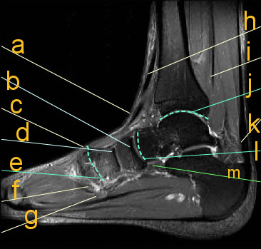
Magnetic Resonance Imaging Mri Image Showing Foot Muscles And Download Scientific Diagram from www.researchgate.net There are 10 intrinsic muscles located in the sole of the foot. The foot contains many bones, muscles, tendons, and other structures. Radiologists perform ankle imaging to assess injuries of the foot and ankle anatomy. You can click the links in the image, or the links below the image to find out more information on any muscle group. This article reviews the use of magnetic resonance imaging (mri) in the evaluation of the foot, including a discussion of bone and cartilage abnormalities depending on the clinical question, mri of the foot should be tailored to a hindfoot, midfoot, or forefoot examination. Extensor brevis and longus muscles. Tendinous, ligamentous, and muscle abnormalities. Structures of the foot shown in this illustration are:
The muscles acting on the foot can be divided into two distinct groups;
If more detail is needed, however, an orthopedic doctor will likely want to do magnetic resonance imaging (mri)—a technique that uses a. Muscles of the lower limb | anatomy model. Attached to the bones of the skeletal system are about 700 named. Almost all muscles cross at least one joint (moveable connection between two bones) and cause an action across that joint. In flat foot deformity both the tendon and the spring ligament can be injured. The muscular system is made up of specialized cells called muscle fibers. They act collectively to stabilise the arches of the foot, and individually to control movement of the digits. Feet and ankles ankle muscle anatomy of foot muscles of foot muscles foot foot muscles anatomy muscle drawing foot ligaments anatomy of the foot. You can click the links in the image, or the links below the image to find out more information on any muscle group. Neuropathies around the elbow joint. Find the best weight lifting exercises that target each muscle or groups of muscles. Leg and foot (exam 2). Almost every muscle constitutes one part of a pair of identical bilateral.
This article reviews the use of magnetic resonance imaging (mri) in the evaluation of the foot, including a discussion of bone and cartilage abnormalities depending on the clinical question, mri of the foot should be tailored to a hindfoot, midfoot, or forefoot examination foot muscles mri. Neuropathies around the elbow joint.